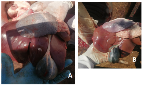
Small ruminants, particularly goats, are essential to the livelihoods of rural communities in Sudan; however, their productivity is threatened by tick-borne diseases such as anaplasmosis. This study aimed to determine the prevalence of anaplasmosis in goats slaughtered in El Daein city, East Darfur State, Sudan, and to assess associated epidemiological and clinical risk factors. A cross-sectional study was conducted from November 2022 to February 2023 in three slaughterhouses. Blood samples (n = 175) were collected from randomly selected goats and examined for Anaplasma spp. using Giemsa-stained blood smears. Ante-mortem and post-mortem examinations were performed to identify clinical and pathological abnormalities. Data were analyzed using chi-square and logistic regression tests. The overall prevalence of anaplasmosis was 62.3%. No statistically significant association (P > 0.05) was observed between infection and sex, age, breed, or post-mortem pathological lesions including splenomegaly, gallbladder enlargement, or jaundice. However, a significant association was detected between infection and pale mucous membranes (P = 0.031), indicating anemia as a likely consequence of erythrocytic infection. Most infections appeared subclinical, suggesting endemic stability in the study area. The high prevalence indicates widespread circulation of Anaplasma among goats in East Darfur, likely driven by favorable ecological conditions for tick proliferation. Strengthening tick control strategies and routine diagnostic surveillance is recommended to reduce disease burden and improve small ruminant productivity in the region.
| Published in | Animal and Veterinary Sciences (Volume 13, Issue 6) |
| DOI | 10.11648/j.avs.20251306.16 |
| Page(s) | 211-215 |
| Creative Commons |
This is an Open Access article, distributed under the terms of the Creative Commons Attribution 4.0 International License (http://creativecommons.org/licenses/by/4.0/), which permits unrestricted use, distribution and reproduction in any medium or format, provided the original work is properly cited. |
| Copyright |
Copyright © The Author(s), 2025. Published by Science Publishing Group |
Anaplasmosis, Goat, Prevalence, Risk Factor
Risk factor | Total no. of goats | Goats positive for Anaplasma | Percentages of goats positive for Anaplasma | Df | X2 | P-value | |
|---|---|---|---|---|---|---|---|
Sex | Male | 59 | 38 | 64.4% | |||
Female | 116 | 71 | 61.2% | 1 | 0.170 | 0.404 | |
Age | less than year | 108 | 66 | 61.1% | |||
1-3 years | 15 | 9 | 65.4% | 2 | 0.309 | 0.857 | |
over 3 years | 52 | 34 | 60.0% | ||||
Gallbladder | Enlargement | 160 | 100 | 62.5% | |||
Normal | 13 | 8 | 61.5% | 2 | 0.135 | 0.935 | |
Empty | 2 | 1 | 50.0% | ||||
Spleen | Splenomegaly | 32 | 18 | 56.3% | |||
Normal | 143 | 91 | 63.6% | 1 | 0.607 | 0.280 | |
Mucous membrane | Pale | 6 | 6 | 100% | |||
Congested | 2 | 0 | 0.00% | ||||
Normal | 167 | 103 | 61.7% | 2 | 6.962 | 0.031* | |
Hair condition | Brittle | 6 | 4 | 66.7% | |||
Normal | 169 | 105 | 62.1% | 1 | .051 | 0.592 | |
EDTA | Ethylenediaminetetraacetic Acid |
Df | Degree of Freedom |
CI | Confidence Interval |
RBCs | Red Blood Cells |
| [1] | Ahmed, M. A., Musa, L. M. A., & Haroun, E. M. (2016). Livestock and livelihoods in Darfur: A regional overview. Sudan Journal of Veterinary Research, 31(2), 45–52. |
| [2] | Ngambi, J. W.; Alabi, O. J.; Noris, D. (2013). Role of goats in food security, poverty alleviation and prosperity with special reference to Sub-Saharan Africa: A review. Indian J. of Anim. Res, 47(1), 1–9. |
| [3] | FAO. (2020). The state of livestock in the developing world. Food and Agriculture Organization of the United Nations, Rome. |
| [4] | Wilson, R. T., (1991). Small ruminant production and the small ruminant genetic resource in tropical Africa (No. 88). Food and Agriculture Organization (FAO). |
| [5] | El Hussein, A. M., Hassan, S. M., and Salih, D. A. (2012): Current situation of tropical theileriosis, bovine babesiosis and anaplasmosis in the Sudan. Parasitology Research. 2012, 111(2): 503-8. |
| [6] | Hartelt, K., Oehme, R., Frank, H., Brockmann, S. O., Hassler, D. and Kimmig, P., (2004). Pathogens and symbionts in ticks: prevalence of Anaplasma phagocytophilum (Ehrlichia sp.), Wolbachia sp., Rickettsia sp., and Babesia sp. in Southern Germany. International Journal of Medical Microbiology. 293 (Suppl. 37), 86–92. |
| [7] | Ewing, S. A., (1981). Transmission of Anaplasma marginale by arthropods. In Proceedings of the 7th National Anaplasmosis Conference. Mississippi State University, Mississippi State (Vol. 395, p. 423). |
| [8] | Al Faki, B. H., (2004). Studies on ticks and tick–borne diseases of export Sheep at Alkadaro Slaughterhouse. PhD thesis, University of Khartoum: Khartoum, Sudan. |
| [9] | Rymaszewska, A., & Grenda, S. (2008). Bacteria of the genus Anaplasma-Characteristics of Anaplasma and their vectors: A review. Veterinary Medicine, 53(11), 573–584. |
| [10] | Kocan, K. M., de la Fuente, J., Guglielmone, A. A., & Meléndez, R. D. (2004). Antigens and alternatives for control of Anaplasma marginale infection in cattle. Clinical Microbiology Reviews 17(4): 698-712. |
| [11] | Thrusfield, M., (2007). Sample size determination. Veterinary epidemiology, 3, pp. 185-189. |
| [12] | Murray, P. R., Baron, E. J., Pfaller, M. A., Tenover, F. C., & Yolken, R. H. (1977). Manual of Clinical Microbiology. American Society for Microbiology Press, Washington, D. C. |
| [13] | Asrar Abdelrady, A. (2017). Prevalence of tick-borne diseases in small ruminants in Sudan. Sudan Journal of Veterinary Science and Animal Husbandry, 56(1), 23–31. |
| [14] | Nasreen, A., Saeed, K., Khan, A., Niaz, S. and Akhtar, N., (2016). Serodiagnosis and haematological effect of anaplasmosis in goats and sheep of district Mardan, Khyber Pakhtunkhwa, Pakistan. World J Zool, 11(2), pp. 67-80. |
| [15] | Masoud Soosaraei, Mousa Motavalli Haghi, Fariborz Etemadifar, Mahdi Fakhar, Saeed Hosseini Teshnizi, Shabnam Asfaram, Bahman Rahimi Esboei. (2020). Status of Anaplasma spp. infection in domestic ruminants from Iran: A systematic review with meta-analysis, Parasite Epidemiology and Control, 11, e00173, |
| [16] |
. Whittier, W. D., Currin, N., & Currin, J. F. (2009). Anaplasmosis in beef cattle (Publication No. 400-465). Virginia Cooperative Extension, Virginia Tech.
https://vtechworks.lib.vt.edu/bitstreams/5d96b41a-f478-42a8-bbd6-dcabe87605de/download |
| [17] | Nabil M. A. M. A (2003). Prevalence of bovine anaplasmosis in Malaysia farms. Faculty of Veterinary Medicine, University of Putra Malysia: Serdang.. |
| [18] | Lee, S; Mossaad, E; Ibrahim, A. M.; Ismail, A. A.; Moumouni, P. F. A.; Liu, M.; Ringo, A. E.; Gao, y.; Guo, H.; Li, J.; Efstratiou, A.; Musinguzi, P.; Tamador Angara, E. E.; Suganuma, K.; Inoue, N.; Xuan, x. (2018). Detection and molecular characterization of tick-borne pathogens infecting sheep and goats in Blue Nile and West Kordofan states in Sudan. Ticks and Tick-borne Diseases. 9 (3): 598-604, |
| [19] | Urquhart, G. M., J. Armour, J. L. Duncan, A. M. Dunn and F. W. Jennings (1996). Veterinary Parasitology. 2 ed. USA: Blackwell Science Incorporated, nd pp: 242-253. |
APA Style
Jibreel, K. A. M., Abdalla, M. A., Elrayah, H. O., Elnaeim, S., Ibrahim, M. T. (2025). Anaplasma Infection in Goats in El Daein, East Darfur State, Sudan: An Epidemiological Study. Animal and Veterinary Sciences, 13(6), 211-215. https://doi.org/10.11648/j.avs.20251306.16
ACS Style
Jibreel, K. A. M.; Abdalla, M. A.; Elrayah, H. O.; Elnaeim, S.; Ibrahim, M. T. Anaplasma Infection in Goats in El Daein, East Darfur State, Sudan: An Epidemiological Study. Anim. Vet. Sci. 2025, 13(6), 211-215. doi: 10.11648/j.avs.20251306.16
AMA Style
Jibreel KAM, Abdalla MA, Elrayah HO, Elnaeim S, Ibrahim MT. Anaplasma Infection in Goats in El Daein, East Darfur State, Sudan: An Epidemiological Study. Anim Vet Sci. 2025;13(6):211-215. doi: 10.11648/j.avs.20251306.16
@article{10.11648/j.avs.20251306.16,
  author = {Khalda Abdelgadire Mohammed Jibreel and Mohamed Abdelsalam Abdalla and Hind Osman Elrayah and Sarra Elnaeim and Mohamed Tajeldin Ibrahim},
  title = {Anaplasma Infection in Goats in El Daein, East Darfur State, Sudan: An Epidemiological Study},
  journal = {Animal and Veterinary Sciences},
  volume = {13},
  number = {6},
  pages = {211-215},
  doi = {10.11648/j.avs.20251306.16},
  url = {https://doi.org/10.11648/j.avs.20251306.16},
  eprint = {https://article.sciencepublishinggroup.com/pdf/10.11648.j.avs.20251306.16},
  abstract = {Small ruminants, particularly goats, are essential to the livelihoods of rural communities in Sudan; however, their productivity is threatened by tick-borne diseases such as anaplasmosis. This study aimed to determine the prevalence of anaplasmosis in goats slaughtered in El Daein city, East Darfur State, Sudan, and to assess associated epidemiological and clinical risk factors. A cross-sectional study was conducted from November 2022 to February 2023 in three slaughterhouses. Blood samples (n = 175) were collected from randomly selected goats and examined for Anaplasma spp. using Giemsa-stained blood smears. Ante-mortem and post-mortem examinations were performed to identify clinical and pathological abnormalities. Data were analyzed using chi-square and logistic regression tests. The overall prevalence of anaplasmosis was 62.3%. No statistically significant association (P > 0.05) was observed between infection and sex, age, breed, or post-mortem pathological lesions including splenomegaly, gallbladder enlargement, or jaundice. However, a significant association was detected between infection and pale mucous membranes (P = 0.031), indicating anemia as a likely consequence of erythrocytic infection. Most infections appeared subclinical, suggesting endemic stability in the study area. The high prevalence indicates widespread circulation of Anaplasma among goats in East Darfur, likely driven by favorable ecological conditions for tick proliferation. Strengthening tick control strategies and routine diagnostic surveillance is recommended to reduce disease burden and improve small ruminant productivity in the region.},
 year = {2025}
}
TY  - JOUR
T1  - Anaplasma Infection in Goats in El Daein, East Darfur State, Sudan: An Epidemiological Study
AU  - Khalda Abdelgadire Mohammed Jibreel
AU  - Mohamed Abdelsalam Abdalla
AU  - Hind Osman Elrayah
AU  - Sarra Elnaeim
AU  - Mohamed Tajeldin Ibrahim
Y1  - 2025/12/26
PY  - 2025
N1  - https://doi.org/10.11648/j.avs.20251306.16
DO  - 10.11648/j.avs.20251306.16
T2  - Animal and Veterinary Sciences
JF  - Animal and Veterinary Sciences
JO  - Animal and Veterinary Sciences
SP  - 211
EP  - 215
PB  - Science Publishing Group
SN  - 2328-5850
UR  - https://doi.org/10.11648/j.avs.20251306.16
AB  - Small ruminants, particularly goats, are essential to the livelihoods of rural communities in Sudan; however, their productivity is threatened by tick-borne diseases such as anaplasmosis. This study aimed to determine the prevalence of anaplasmosis in goats slaughtered in El Daein city, East Darfur State, Sudan, and to assess associated epidemiological and clinical risk factors. A cross-sectional study was conducted from November 2022 to February 2023 in three slaughterhouses. Blood samples (n = 175) were collected from randomly selected goats and examined for Anaplasma spp. using Giemsa-stained blood smears. Ante-mortem and post-mortem examinations were performed to identify clinical and pathological abnormalities. Data were analyzed using chi-square and logistic regression tests. The overall prevalence of anaplasmosis was 62.3%. No statistically significant association (P > 0.05) was observed between infection and sex, age, breed, or post-mortem pathological lesions including splenomegaly, gallbladder enlargement, or jaundice. However, a significant association was detected between infection and pale mucous membranes (P = 0.031), indicating anemia as a likely consequence of erythrocytic infection. Most infections appeared subclinical, suggesting endemic stability in the study area. The high prevalence indicates widespread circulation of Anaplasma among goats in East Darfur, likely driven by favorable ecological conditions for tick proliferation. Strengthening tick control strategies and routine diagnostic surveillance is recommended to reduce disease burden and improve small ruminant productivity in the region.
VL  - 13
IS  - 6
ER  -
Department of Preventive Medicine and Public Health, Sudan University of Science and Technology, Hilat Koukou, Sudan
Department of Preventive Medicine and Public Health, Sudan University of Science and Technology, Hilat Koukou, Sudan
Department of Preventive Medicine and Public Health, Sudan University of Science and Technology, Hilat Koukou, Sudan
Department of Preventive Medicine and Public Health, Sudan University of Science and Technology, Hilat Koukou, Sudan
Department of Animal Production, Sudan University of Science and Technology, Hilat Koukou, Sudan